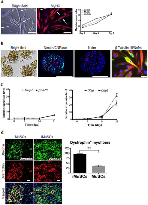
| In vitro multipotent differentiation assays showed that iMuSCs were able to fuse with MyHC+ (Myosin heavy chain) myotubes in muscle differentiation medium with a similar fusion index as control MuSCs and C2C12 myoblasts (Fig. 2a). The iMuSCs were also capable of differentiating into osteogenic lineages (Supplementary Fig. S2) within osteogenic medium with BMP2. The iMuSCs could also be easily and effectively induced into a neurogenic lineage via neurosphere formation once cultured in neural stem cell medium (see Method) for one week (Fig. 2b), whereas the control primary myoblasts and MuSCs showed no sign of forming these structures. The iMuSCs-induced neurospheres exhibited a neural phenotype and expressed Nestin, CNPase and Nefm (Neurofilament) (Fig. 2b). After three weeks, the neurospheres when re-plated in a laminin/polyornithine coated monolayer culture in neural differentiation medium, could differentiate into the three major neural lineages (neurons, astrocytes, and oligodendrocytes) and they expressed Mtap2, β-Tubulin III, Nefm, Nestin and Olig1/2 (Oligodendrocyte transcription factor 1/2) (Fig. 2b,c). To further investigate the origin of the iMuSCs, we performed in vivointramuscular transplantation studies. Equal numbers of iMuSCs and control MuSCs were injected into the TA muscles of six 6–8 week-old male mdx/SCID mice (Jackson Lab, USA). Two and three weeks after cell implantation, we detected Utrophin and Dystrophin (Fig. 2d) expression in the host TA muscles, and observed that the iMuSCs formed larger and more robust Dystrophin+ muscle grafts compared to the control MuSCs (Fig. 2d). |