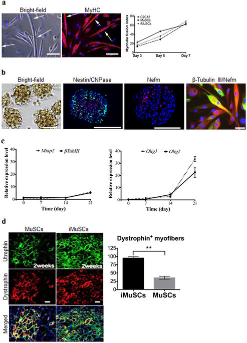
| (a) Induced myotube formation of iMuSCs. Myotubes expressed MyHC (red). The fusion index was similar to the control C2C12 and MuSCs. (b) Representative bright-field picture shows iMuSCs-formed neurospheres floating in suspension. Immunofluorescence staining of cryosectioned neurospheres shows Nestin (green), CNPase (red), and Nefm (red) positive cells. Plated 21 day differentiated neurospheres in ND medium show neural phenotype; β-Tubulin III (red), and Nefm (green). Nuclei were stained with DAPI. Scale bar = 10 and 100 μm. (c) Gene expression kinetics of Mtap2 and β-Tubulin III, and Olig1 and Olig2 in the neural differentiating iMuSCs analyzed by qPCR. Data were compared to undifferentiated iMuSCs, and are presented as the mean ±SEM of 5 biological replicates. (d) Engraftment of iMuSCs after intramuscular cell implantation. IF staining shows Utrophin+ (green) and Dystrophin+ (red) muscle engraftment of control MuSCs and iMuSCs in mdx/scid mice 2 weeks after cell injection. Scale bar = 100 μm. Quantification of Dystrophin+ myofibers. **P < 0.01. |