President Trump’s inauguration speech

トランプ大統領の就任演説の全文を掲載しておこう。
*訳文だと、訳者の思惑や感情が刷り込まれている、或いは意図的に意訳している可能性があるので、原文を読む必要がある。ましてやTVのニュース解説なんて言わずもがなだ。
*ちょっとだけ私見を。どうも「虐げと回復」がテーマのように見える。どっかで見たこと聞いたことがあるよな、ないような・・。
 

Chief Justice Roberts, President Carter, President Clinton, President Bush, President Obama, fellow Americans, and people of the world:

thank you.

We, the citizens of America, are now joined in a great national effort to rebuild our country and to restore its promise for all of our people.

Together, we will determine the course of America and the world for years to come.

We will face challenges. We will confront hardships. But we will get the job done.Every four years, we gather on these steps to carry out the orderly and peaceful transfer of power, and we are grateful to President Obama and First Lady Michelle Obama for their gracious aid throughout this transition. They have been magnificent.

Today’s ceremony, however, has very special meaning. Because today we are not merely transferring power from one Administration to another,or from one party to another – but we are transferring power from Washington, D.C. and giving it back to you, the American People.

For too long, a small group in our nation’s Capital has reaped the rewards of government while the people have borne the cost.

Washington flourished-but the people did not share in its wealth.Politicians prospered – but the jobs left, and the factories closed.The establishment protected itself, but not the citizens of our country.

Their victories have not been your victories; their triumphs have notbeen your triumphs; and while they celebrated in our nation’s Capital,there was little to celebrate for struggling families all across our land.

That all changes – starting right here, and right now, because this moment is your moment: it belongs to you.

It belongs to everyone gathered here today and everyone watching all across America.This is your day. This is your celebration.

And this, the United States of America, is your country. What truly matters is not which party controls our government, but whether our government is controlled by the people.

January 20th 2017, will be remembered as the day the people became the rulers of this nation again.The forgotten men and women of our country will be forgotten no longer.

Everyone is listening to you now.You came by the tens of millions to become part of a historic movement the likes of which the world has never seen before. At the center of this movement is a crucial conviction: that a nation exists to serve its citizens.

Americans want great schools for their children, safe neighborhoods for their families, and good jobs for themselves.These are the just and reasonable demands of a righteous public.

But for too many of our citizens, a different reality exists: Mothers and children trapped in poverty in our inner cities; rusted-out factories scattered like tombstones across the landscape of our nation; an education system, flush with cash, but which leaves our young and beautiful students deprived of knowledge; and the crime and gangs and drugs that have stolen too many lives and robbed our country of so much unrealized potential.

This American carnage stops right here and stops right now. We are one nation – and their pain is our pain. Their dreams are our dreams; and their success will be our success. We share one heart,one home, and one glorious destiny.

The oath of office I take today is an oath of allegiance to all Americans. For many decades, we’ve enriched foreign industry at the expense of American industry; Subsidized the armies of other countries while allowing for the very sad depletion of our military; We’ve defended other nation’s borders while refusing to defend our own; And spent trillions of dollars overseas while America’s infrastructure has fallen into disrepair and decay.

We’ve made other countries rich while the wealth, strength, and confidence of our country has disappeared over the horizon. One by one, the factories shuttered and left our shores, with not even a thought about the millions upon millions of American workers left behind.

The wealth of our middle class has been ripped from their homes and then redistributed across the entire world. But that is the past. And now we are looking only to the future. We assembled here today are issuing a new decree to be heard in every city, in every foreign capital, and in every hall of power. From this day forward, a new vision will govern our land.

From this moment on, it’s going to be America First. Every decision on trade, on taxes, on immigration, on foreign affairs, will be made to benefit American workers and American families.We must protect our borders from the ravages of other countries making our products, stealing our companies, and destroying our jobs. Protection will lead to great prosperity and strength. I will fight for you with every breath in my body – and I will never,ever let you down.

America will start winning again, winning like never before.We will bring back our jobs. We will bring back our borders. We will bring back our wealth. And we will bring back our dreams.We will build new roads, and highways, and bridges, and airports, and tunnels, and railways all across our wonderful nation.

We will get our people off of welfare and back to work – rebuilding our country with American hands and American labor. We will follow two simple rules: Buy American and Hire American.We will seek friendship and goodwill with the nations of the world -but we do so with the understanding that it is the right of all nations to put their own interests first.

We do not seek to impose our way of life on anyone, but rather to let it shine as an example for everyone to follow.

We will reinforce old alliances and form new ones – and unite the civilized world against Radical Islamic Terrorism, which we will eradicate completely from the face of the Earth.

At the bedrock of our politics will be a total allegiance to the United States of America, and through our loyalty to our country, we will rediscover our loyalty to each other. When you open your heart to patriotism, there is no room for prejudice.

The Bible tells us, “how good and pleasant it is when God’s people

live together in unity.” We must speak our minds openly, debate our disagreements honestly, but always pursue solidarity. When America is united, America is totally unstoppable.

There should be no fear – we are protected, and we will always be protected. We will be protected by the great men and women of our military and law enforcement and, most importantly, we are protected by God.

Finally, we must think big and dream even bigger. In America, we understand that a nation is only living as long as it is striving. We will no longer accept politicians who are all talk and no action -constantly complaining but never doing anything about it. The time for empty talk is over.

Now arrives the hour of action. Do not let anyone tell you it cannot be done. No challenge can match the heart and fight and spirit of America. We will not fail. Our country will thrive and prosper again.We stand at the birth of a new millennium, ready to unlock the mysteries of space, to free the Earth from the miseries of disease,and to harness the energies, industries and technologies of tomorrow. A new national pride will stir our souls, lift our sights, and heal our divisions.

It is time to remember that old wisdom our soldiers will never forget: that whether we are black or brown or white, we all bleed the same red blood of patriots, we all enjoy the same glorious freedoms, and we all salute the same great American Flag.And whether a child is born in the urban sprawl of Detroit or the windswept plains of Nebraska, they look up at the same night sky, they fill their heart with the same dreams, and they are infused with the breath of life by the same almighty Creator.

So to all Americans, in every city near and far, small and large, from mountain to mountain, and from ocean to ocean, hear these words: You will never be ignored again.Your voice, your hopes, and your dreams, will define our American destiny. And your courage and goodness and love will forever guide us along the way.

Together, We Will Make America Strong Again.We Will Make America Wealthy Again.We Will Make America Proud Again.We Will Make America Safe Again. And, Yes, Together, We Will Make America Great Again. Thank you, God Bless You, And God Bless America.

カテゴリー: General | President Trump’s inauguration speech はコメントを受け付けていません

VR Roller Coaster 360° VIDEO

左クリックを保持したままグリグリ回すと、360度の画面が見れるぞ!

カテゴリー: General | VR Roller Coaster 360° VIDEO はコメントを受け付けていません

謹賀新年

                                                昨年は大変お世話になりました。
                                               今年も宜しくお願い申し上げます。
カテゴリー: General | 謹賀新年 はコメントを受け付けていません

Beautiful bird singing

 よく眠れそうだ。。。
カテゴリー: General | Beautiful bird singing はコメントを受け付けていません

treatment? for insomnia

夜中の3時ごろ眼が醒めてしまい、その後寝付けないことがある。そんな時、上のような睡眠導入動画を試してみる。これまでは、この手の動画を聴いても全く効果がなかった。確かに穏やかな音楽がチョイスされてはいる。だが、高音領域の音が多いので、場合によってはキンキン響いて耳障りなことがある。それらと違って、この音楽は低音がはっきりと聴こえるよう工夫されている。この低音域、つまり副旋律が適度なボリュームを持つ構成となっている。そして、この副旋律が不快で聴きたくない思い出したくない領域の音を遮蔽しているように思われる。このノイズ遮断様効果によって、高音域の鳥のさえずりが冴えわたり、大変心地よく耳に響いてくる。それで、ついうとうとしてしまう。まあ、1個人の意見ではあるが。

耳鳴りの治療方法の一つに音響療法というのがある。原理はよく似ている。音響療法の要点についてについてはこのサイトに分かりやすく書かれている(→音響効果)。参考までに。

カテゴリー: General | treatment? for insomnia はコメントを受け付けていません

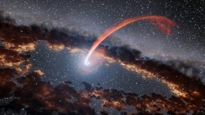
Supermassive Black Hole Eating That Star

 これはすごい!良く撮影できたもんだ!
pia20027_updated
 →こちらの記事
翻訳と解説はこちら(→ブラックホールが星を捕食!
この日本語解説にある動画がまた実に面白い。まるでフィクション漫画を見ているようだ。
カテゴリー: 宇宙 | Supermassive Black Hole Eating That Star はコメントを受け付けていません

Your Japanese is good?

確信犯・奇特…日本語、正しく使えますか? 文化庁調査

文化庁の2015年度国語世論調査で、「日本語を大切にしている」と答えた人は8割弱にのぼることが明らかになった。その一方で、慣用句の調査では、「確信犯」「琴線に触れる」を本来の意味で使う人は少なかった。

毎日使う日本語について、「大切にしていると思う」は34・9%、「あまり意識したことはないが、考えてみれば大切にしていると思う」は43・6%で、計78・5%に達した。01年度調査から1割近く増えた。

また、敬語について、「伝統的な美しい日本語として、豊かな表現が大切にされるべきだ」と答えた人も64・1%に上り、「新しい時代にふさわしく、簡単でわかりやすいものであるべきだ」とする26・1%を大きく上回った。ほぼ互角だった97年度調査(前者が46・9%、後者が41・4%)と比べ、敬語を重視する傾向が浮き彫りになった。

「確信犯」の意味は、「政治的・宗教的などの信念に基づいて正しいと信じてなされる行為(その行為を行う人)」だが、その意味と回答したのはわずか17%で、7割は「悪いことだとわかっていながらなされる行為(その行為を行う人)」の意味だと答えた。

守真弓 2016年9月22日11時16分

・・・
>「日本語を大切にしている」と答えた人は8割弱にのぼる

大変良いことだと思うが、「日本語を大切にしている」と答えた人が100%というアンケート結果になって欲しかった。さらに、「ちゃんとした日本語をしゃべっている」かという質問もして欲しかった。「ちゃんとした日本語をしゃべっている」人が8割弱なら、おそらく「日本語を大切にしている」と答える人は10割になるに違いない。実際、ちゃんとした日本語をしゃべれる人は実に少ない。

>「新しい時代にふさわしく、簡単でわかりやすいものであるべきだ」

あらかじめ用意されていた解答ではないかとguessするが、敬語に関する答えのひとつとしてはトンチンカンではないかと思う。敬語の文法はすでに確立されていて、どうにも変えようがないのだ。ちゃんとした日本語をしゃべれない人たちの敵意からくる感情ではないかとつい勘ぐってしまう。

>「確信犯」の意味を「悪いことだとわかっていながらなされる行為(その行為を行う人)」と答えた人が7割・・・

小生もそう思っていた。「確信犯」とは 「政治的・宗教的などの信念を知りながら、意図的にその信念に反する行為を行なう人」じゃないの?

早速ネットで調べて見ると、確かに小生の定義は、確信犯ではなくて故意犯ではないかと思われる。しかし、「新しい時代にふさわしく、簡単でわかりやすいものであるべき」というのなら、小生の定義でもいいのではないだろうか。

最後のパラグラフのような末節的な議論をする前に、まずはちゃんとした日本語をしゃべれるようにしたい。これができないで、小学校からの英語教育なんてちゃんちゃらおかしい。学校関係者諸氏にお願いしたい。情操教育や英語教育はさておいて、読み書きソロバンをしっかり教えて欲しい。

カテゴリー: 教育, 言語 | Your Japanese is good? はコメントを受け付けていません

Tour 2016 in Hokkaido

DSC_001


DSC_003 (1)


DSC_003 (2)


DSC_004


DSC_005
 今年も北海道! 台風の影響はほぼなかった...
カテゴリー: General | Tour 2016 in Hokkaido はコメントを受け付けていません

iMuSCs, #That’s it

STAP細胞とiMuSCsおよびMuse細胞との決定的な違いはその作成方法にある。

先にも書いたが、STAP細胞はin vitroによって作成され、iMuSCsおよびMuse細胞はin vivoにおいて誘導される多能性幹細胞のようだ。

細胞の調達という点からすれば、STAP細胞の方がはるかに有用だ。何故なら、生体を傷害したり、炎症反応を起こしたりする手間が要らないから。しかも、簡単に採取できる血中リンパ球などから作成できるのでなおさら良い。作成法も極めて簡単で、得られたリンパ球を弱酸処理するだけ。

さて、iMuSCs論文において、著者らは傷害部位局所において多能性幹細胞が発生したと力説するが、造血幹細胞が血流を介して迷入したというストーリーがどうしても否定できない。また、傷害筋組織中に多能性幹細胞が発生するという話題提供だけで、iMuSCsの応用など発展性に欠けているのも問題だ。

STAP細胞作成方法には、弱酸処理だけでなく他にもいろいろあるようだ。いずれにせよ、STAP現象をパーフェクトに証明できる優秀な研究者の登場を期待したい。

カテゴリー: General | iMuSCs, #That’s it はコメントを受け付けていません

iMuSCs, #13b

次にiMuSCsの作成方法とSTAP細胞の作成方法を比較・検討しておく。

iMuSCsの作成方法はすでに説明した。STAP細胞の作成方法については、小保方氏のNature論文に載っている。次の箇所だ。

Stimulus-triggered fate conversion of somatic cells into pluripotencyのMethodsより引用
Tissue collection and low-pH treatment
To isolate CD45+ haematopoietic cells, spleens were excised from 1-week-old Oct4-gfp mice (unless specified otherwise), minced by scissors and mechanically dissociated with pasture pipettes. Dissociated spleen cells were suspended with PBS and strained through a cell strainer (BD Biosciences). After centrifuge at 1,000 r.p.m. for 5 min, collected cells were re-suspended in DMEM medium and added to the same volume of lympholyte (Cedarlane), then centrifuged at 1,000g for 20 min. The lymphocyte layer was taken out and stained with CD45 antibody (ab25603, Abcam). CD45-positive cells were sorted by FACS Aria (BD Biosciences). After cell sorting, 1 × 106 CD45-positive cells were treated with 500 μl of low-pH HBSS solution (titrated to pH5.7 by HCl) for 25 min at 37 °C, and then centrifuged at 1,000 r.p.m. at room temperature for 5 min. After the supernatant (low-pH solution) was removed, precipitated cells were re-suspended and plated onto non-adhesive culture plates (typically, 1 × 105 cells ml−1) in DMEM/F12 medium supplemented with 1,000 U LIF (Sigma) and 2% B27 (Invitrogen). Cell cluster formation was more sensitive to the plating cell density than the percentage of Oct4-GFP+ cells. The number of surviving cells was sensitive to the age of donor mice and was low under the treatment conditions above when adult spleens were used. The addition of LIF during days 2–7 was essential for generating Oct4-GFP+ STAP cell clusters on day 7, as shown in Extended Data Fig. 1f. Even in the absence of LIF, Oct4-GFP+ cells (most of them were dim in signal) appeared transiently during days 2–5 in culture of low-pH-treated CD45+ cells, but subsequently disappeared, indicating that there is a LIF-independent early phase, whereas the subsequent phase is LIF-dependent.
組織収集と低酸性処理
CD45陽性造血細胞を分離するため、1週齢のOct4-gfpマウス(他の箇所で特に説明がなければ)から得られた脾臓をハサミで細かく刻み、パスツールピペットで機械的に細断した。細断した脾細胞をPBSに浮遊させ、細胞ストレーナー(濾し器、BD Biosciences)を通して濾した。1000回転で5分遠心後、回収した細胞はDMEM培養液に浮遊し、同量のリンパ球浮遊液をリンパ球分離液(Cedarlane)にアプライして、1000で20分間遠心した。リンパ球層を回収し、CD45抗体で染色した。CD45陽性細胞をFACS Aria (BD Biosciences)にて分離した。分離後、1×10⁶個のCD45陽性細胞を500μlの低酸性HBSS溶液(塩酸でpH5.7まで希釈)を用い37度で25分間処理した。上清(低酸性液)を除去後、細胞ペレットを1,000 UのLIF(白血病細胞増殖抑制因子)(Sigma)と2% B27 (Invitrogen)を加えたDMEM/F12培養液に再浮遊させ、非付着用培養皿に撒いた。細胞集塊形成は、Oct4-GFP陽性細胞の陽性率よりも撒かれた細胞濃度に依存していた。生存細胞数は、使用されたマウスの週齢に依存し、週齢の進んだマウスを使用した時には同一処理条件下で低値であった。追加データ図1fに見られるように、培養7日目にOct4-GFP陽性STAP細胞を得るため、培養第2日目から7日目の間にLIF(白血病細胞増殖抑制因子)を加える必要があった。LIFの非存在下においても、Oct4-GFP陽性細胞(多くは弱いシグナルであった)は、低酸性処理CD45陽性細胞の培養2日目から5日目の間に一時的に出現し、ほどなく消失した。このことは、培養早期にLIF非依存性の時期があることを示している。一方、これに続く時期はLIF依存性の時期である。
*注:意味を類推して訳出した箇所がある。
つまり、iMuSCsはin vivo(生体内)で発生した細胞集団であり、一方、小保方氏がSTAP細胞と称する細胞集団はin vitro(試験管内)において作成された細胞集団であることが分かる。従って、iMuSCsは、その起源という点からすると、STAP細胞とは全く異なる細胞群であり、むしろin vivo由来という点において、Muse細胞に近い細胞群と言えよう。

さて、上の組織収集と低酸性処理方法において、培養7日目にOct4-GFP陽性STAP細胞を得るため、培養第2日目から7日目の間にLIF・・云々と書かれている箇所がある。このことから、他にも指摘があるように、小保方女史はOct4-GFP陽性細胞をSTAP細胞と勘違いしている可能性がある。だから、記者会見の場で、私は200回もSTAP細胞を作成することに成功したと言ったのではないだろうか。

ある細胞が多能性幹細胞であるためには、言い換えるとOct4-GFP陽性細胞がSTAP細胞であるためには、Oct4-GFP陽性細胞が自発的に個体へと成長することを証明する必要がある。


カテゴリー: 最新医学知識 | iMuSCs, #13b はコメントを受け付けていません

iMuSCs, #13a

まず始めに、この論文を読んで改めて生命の素晴らしさに感服する。非常に複雑であるにもかかわらず、見事なほどに緻密な自律制御から成り立っているのが生命だ。生命と非生命との決定的な違いは、この自律性にある。生命現象は、自律性という自己制御を介して、最終的にひとつの成熟した生命集合体である個体へと成長を遂げる。一方、非生命は人の手による自動制御を行なうことはできるが、分化増殖して最終的にひとつの生命体へと変化する自律性はない。

さて、話を戻そう。

当論文において著者らが行なった実験は非常に単純で分かりやすい。抄録に書いてある通りだ。

まず、傷害誘導性筋細胞由来幹細胞作成方法だが、マウスの前脛骨筋を傷つける。本論にはlaceration injuryと書いてあるので、ナイフか何かで切り付けたのだろう。それから、前脛骨筋を(恐らく、図にあるとおり、筋1本まるごと)採取する。4日培養後に得られた細胞の一部が傷害誘導性筋細胞由来幹細胞(以下、iMuSCs)というわけだ。

ここで、彼らが優れている最初のポイントは、iMuSCsの分離培養技術だ。図からするとESC培養液とは、ESGRO Complete PLUS Clonal Grade Medium (Millipore, USA)のことだと思うが、この培養液に出会うまで相当の日数を要したはずだ。また、全筋細胞のおよそ0.1%というから、iMuSCsを確実に得るために、これまた相当の試行錯誤があったのではないだろうか。この知識と技術がいわゆる伝統芸(技術)というものだ。

アメリカに限らず時代をリードしているラボ(例えば、山中教授の所属する京都大学IPS研究所)は、このような伝統的かつ独創性豊かな技術と知識が満ち溢れている。これがやる気満々の若い研究者を引き付けるのだ。必要な時には、この技術や知恵を交換し合ってコラボすることがある。当たり前だが、このような伝統技術を外部に漏らすことは固く禁じられている。

次に、図aのmusle growth mediumについて簡単にコメントしておく。本論を読むと、この培養液は、10%馬血清(HS)1%鶏胎抽出液(CEE、Accurate Chemical Co., UK)1%ペニシリン-ストレプトマイシンを添加したDulbecco’s Modified Eagle’s Medium (DMEM)のこと。この培養液の基本はDMEMだ。DMEMは、通常組織培養に使用する。musle growth mediumとは、これに10%馬血清と1%鶏胎抽出液を加えた培養液だ。なぜDMEMにこれら2つを混ぜると、筋肉の成長が促進されるのか不明であるが、良くさぐり当てたものだと関心する。

カテゴリー: General | iMuSCs, #13a はコメントを受け付けていません

iMuSCs, #12

当論文の所感と簡単な解説を書く前に、大変有用な情報があるので、まずは伝えておこう。
小保方女史らのNature論文が削除されずに、未だに掲載されたままになっている。
これらの論文を撤回した理由も同時に掲載されているので注目したい。
Stimulus-triggered fate conversion of somatic cells into pluripotency

Haruko Obokata, Teruhiko Wakayama, Yoshiki Sasai, Koji Kojima, Martin P. Vacanti, Hitoshi Niwa, Masayuki Yamato & Charles A. Vacanti

Bidirectional developmental potential in reprogrammed cells with acquired pluripotency

Haruko Obokata, Yoshiki Sasai, Hitoshi Niwa, Mitsutaka Kadota, Munazah Andrabi, Nozomu Takata, Mikiko Tokoro, Yukari Terashita, Shigenobu Yonemura, Charles A. Vacanti & Teruhiko Wakayama

(注)下に図4の翻訳を追加した。
カテゴリー: 最新医学知識 | iMuSCs, #12 はコメントを受け付けていません

iMuSCs, #11

テーブル 1:マウスES細胞とマウス傷害誘導性筋細胞由来幹細胞の共通点と相違点

In vitro 分類 マウスES細胞 傷害誘導性筋細胞由来幹細胞
未分化状態での形態 高いNC比を有する円形細胞 高いNC比を有する円形細胞
単層状態単細胞で成長可能 単層状態単細胞で成長可能
粘性弾性特性 粘性、分化途上で変化 ?
自己複製 する する
表面発現マーカー SSEA-1, c-kit, LIFR SSEA-1, Sca-1, CXCR-4, CD-34
アルフォス活性 高い 高い
テロメラーゼ活性 高いmTRT発現 ?
幹細胞性”遺伝子発現 Oct-3/4, Nanog, Sox-2, Klf-2, Klf-5, Klf-4, GDF-3, Rex-1, Ecat-1, Stat-3, Fox-D3, Vasa, Shall-4, Dax-1, Esrrb, Esg-1, Tbx-3, Tcl-1, Rif-2, Nac-1, Zfp-281, Dppa-3 Oct-3/4, Nanog, Sox-2, Rex-1, Klf-2, Klf-5, Ecat-1, Esrrb, Tbx-3, Blimp-1, c-Met, Fragilis, Ki67, Mrf-4, Msx-1, Myf-5, MyoD, Nesitn, Pax-3, Pax-7, Spry-1
遺伝上/遺伝周辺特性 Oct-3/4, Rex-1, Nanog プロモーターの脱メチル化 ?
遺伝子安定性 安定、核型を維持 安定、二倍体:第5染色体トリソミー への移行性高
2価クロマチン構造 あり ?
胚葉体 あり あり
外胚葉分化 β-Tubulin-III, Mtap-2, Ncam-1, Nestin, Pax-6, Sox-1, Gfap, Olig-1, Olig-2 β-Tubulin-III, Mtap-2, Nestin, Olig-1, Olig-2
内胚葉分化 Bmp-4, Cytokeratins, Hnf-4, Somatostatin Afp
中胚葉分化 α-Cardiac actin, Gata-4, Brachyury, Myh-6/7, Nppa, Myf-5, MyoD Brachyury, α-Smooth muscle actin, Myh-6/7, Desmin, Myogenin
奇形腫形成 あり あり
キメラ形成 あり あり/なし


カテゴリー: 最新医学知識 | iMuSCs, #11 はコメントを受け付けていません

iMuSCs, #10(4)

Figure 4
Skeletal muscle injury induced iMuSCs fulfil several in vivo criteria of pluripotency.
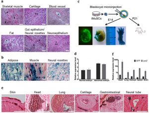
(a) Teratoma formation assay of iMuSCs. Hematoxylin and eosin staining showed differentiated structures of all three germ layers: skeletal muscle (mesoderm, arrows), cartilage (mesoderm, asterisks), blood vessel (mesoderm, asterisk), fat (mesoderm, asterisk), gut epithelium (endoderm, asterisk), neural rosettes (ectoderm, arrow), and neuroepithelium (ectoderm, asterisks) within the same sample. (b) LacZ staining indicated LacZ pre-labelled iMuSC-differentiated structures of adipose, muscle, and neural rosettes contain β-gal+ signals (dark blue dots). Scale bar = 100 μm. (c) Contribution of iMuSCs to mouse embryonic development. Embryos at E14 were analyzed by GFP and LacZ staining, (d) and by qPCR analysis of GFP and LacZ marker gene expression in normally and abnormally developed embryos. (e) The E14 embryos were sectioned and stained with anti-GFP antibody (dark purple). Cells were counterstained with eosin (pink): skin and under-skin, heart, lung, cartilage, gastrointestinal tract, and neural tube. Scale bar = 200 μm. (f) qPCR analysis of GFP and LacZ marker gene expression in the born P21 white pups. Data were pulled together from 6 pups.
 srep17355-f4
 図4
 骨格筋傷害により誘導されたiMuSCsは、in vitroにおいて多能性の基準のいくつかを満たす。
 (a) iMuSCsの奇形腫形成実験。HE染色は、全ての3胚葉層の分化構造を示した:骨格筋(中胚葉、矢印)、軟骨(中胚葉、矢印)、血管(中胚葉、星印)、脂肪(中胚葉、星印)、腸上皮(内胚葉、星印)、神経ロゼッテ(外胚葉、矢印)、神経上皮(外胚葉、矢印)、同一サンプルによる。(b) lacZ染色は、lacZで予めラベルされたたiMuSCにおいて、脂肪組織、筋、神経ロゼッテといった分化構造がβ-gal陽性シグナル(濃紺ドット)を含んでいることを示す。Scale bar = 100 μm。(c)iMuSCsの胚発生に対する関与。E14における胚をGFP及びLacZ染色により解析した。また、(d)定量リアルタイムPCR(qPCR)によって、正常または異常発育した胚におけるGFP及びLacZマーカー遺伝子発現を検討した。(e)E14胚を切片にして、抗GFP抗体(濃紫)で染色した。細胞をエオジン(ピンク)対比染色した:皮膚、皮下組織、心臓、肺、軟骨、胃及び腸、そして神経管。Scale bar = 200 μm。(f)新生P21白色マウスにおけるGFP及びLacZマーカー遺伝子発現に関する量的リアルタイムPCR(qPCR)解析」。データは6匹の新生マウスから得た。
カテゴリー: 最新医学知識 | iMuSCs, #10(4) はコメントを受け付けていません

iMuSCs, #10(3)

Figure 3
Skeletal muscle injury induced iMuSCs fulfil several in vitro criteria of pluripotency.
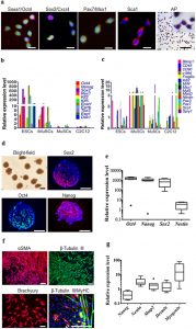
(a) Representative immunofluorescent images of iMuSCs expressing Ssea1 (red), Oct4 (green), Sox2 (red), Cxcr4 (green), Pax7 (red), Msx1 (green), Sca1 (red), and alkaline phosphatase. Nuclei were stained with DAPI (blue). Scale bars = 10 and 200 μm. (b) qPCR analysis of iMuSCs for pluripotent marker genes and (c) myogenic marker genes. Data are represented as the mean ±SEM of 5 biological replicates. (d) Bright-field image of formed embryoid bodies (EBs) in suspension, and immunofluorescence staining of cryosectioned EBs. EBs contained cells positive for Sox2 (red), Oct4 (green), and Nanog (red). Nuclei were stained with DAPI (blue). Scale bar = 100 μm. (e) qPCR data showed upregulation of Oct4, Nanog, Sox2, and no change of Nestin expression compared to control undifferentiated iMuSCs. (f) Immunofluorescence images of differentiated EBs expressing αSMA (red), β-Tubulin III (green), Brachyury (red), and β-Tubulin III β (red) with MyHC (green). Nuclei were stained with DAPI (blue). Scale bar = 100 μm. (g) qPCR data of 7 days differentiated EBs show Nestin, Mtap2, Desmin, and Myogenin upregulation, and Nanog downregulation compared to control undifferentiated iMuSCs. All qPCR data was analyzed by the delta-delta Ct method, and are represented as the mean ±SEM of 5 biological replicates.
srep17355-f3
図3
骨格筋傷害により誘導されたiMuSCsは、in vitroにおいて多能性の基準のいくつかを満たす。
(a)iMuSCsの代表的な粘液蛍光染色像、Ssea1(赤)、Oct4(緑)、Sox2(赤)、Cxcr4(緑)、Pax7(赤)、Msx1(緑)、Sca1(赤)及びアルカリフォスファターゼ。核は DAPI(青)で染色した。スケールバー= 10 and 200 μm。(b) タノウ性マーカー遺伝子に関するiMuSCsのqPCR解析、及び (c) 筋源性マーカー遺伝子。データは、5回反復実験の平均±SEMで示した。(d) 浮遊液中に形成された胚様体(EB)の明視野画像、及び凍結切片した胚様体(EB)の免疫蛍光染色像。EBsはSox2(赤)、 Oct4(緑)、Nanog(赤)に染まる細胞を含んでいる。核は DAPI(青)で染色した。スケールバー= 100 μm。(e) qPCRデータは、Oct4、Nanog、Sox2の発現増加を示した。未分化状態の対照群iMuSCsとの比較において、 Nestinの発現は変化がなかった。(f) 分化したEBsの免疫蛍光染色像:αSMA(赤)、β-Tubulin III (緑)、Brachyury(赤)、β-Tubulin III β (赤)兼MyHC(緑)。核は DAPI(青)で染色した。スケールバー= 100 μm。(g) 分化誘導7日目のEBsのqPCRデータは、Nestin, Mtap2、Desmin、及びMyogeninの発現増強を示した。一方、未分化iMuSCsとの比較において、Nanogは発現の低下を示した。全qPCRデータは、delta-delta Ct 法によって解析され、5回反復実験の平均±SEMで示した。


カテゴリー: 最新医学知識 | iMuSCs, #10(3) はコメントを受け付けていません

iMuSCs, #10(2)

Figure 2
Multiple differentiation and muscle engraftment of iMuSCs.
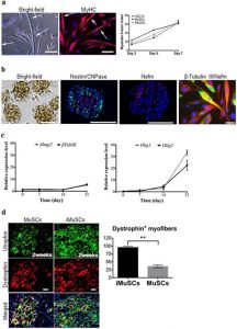
(a) Induced myotube formation of iMuSCs. Myotubes expressed MyHC (red). The fusion index was similar to the control C2C12 and MuSCs. (b) Representative bright-field picture shows iMuSCs-formed neurospheres floating in suspension. Immunofluorescence staining of cryosectioned neurospheres shows Nestin (green), CNPase (red), and Nefm (red) positive cells. Plated 21 day differentiated neurospheres in ND medium show neural phenotype; β-Tubulin III (red), and Nefm (green). Nuclei were stained with DAPI. Scale bar = 10 and 100 μm. (c) Gene expression kinetics of Mtap2 and β-Tubulin III, and Olig1 and Olig2 in the neural differentiating iMuSCs analyzed by qPCR. Data were compared to undifferentiated iMuSCs, and are presented as the mean ±SEM of 5 biological replicates. (d) Engraftment of iMuSCs after intramuscular cell implantation. IF staining shows Utrophin+ (green) and Dystrophin+ (red) muscle engraftment of control MuSCs and iMuSCs in mdx/scid mice 2 weeks after cell injection. Scale bar = 100 μm. Quantification of Dystrophin+ myofibers. **P < 0.01.
srep17355-f2
図2
iMuSCsの多能性と筋移植
(a)iMuSCsによる誘導筋管形成。筋管はMyHC(赤)を発現した。フュージョンインデックスは、コントロールとして用いたC2C12及びMuSCsと類似していた。(b)典型的な明視野を有する写真は、浮遊液中に浮いているiMuSCsによって形成された神経球。凍結切片により作成された神経球の免疫蛍光染色は、Nestin(緑)、CNPase (赤)、Nefm (赤)陽性細胞を示す。ND培養液で21日間培養した神経球は神経表現型を示した:β-Tubulin III(赤)、Nefm (緑)。核は DAPIによって染色された。Scale bar = 10 and 100 μm。(c) 神経系への分化を示すiMuSCsにおけるMtap2、β-Tubulin III、 Olig1、Olig2の遺伝子発現動態。qPCRにより解析した。データは、未分化iMuSCsと比較し、5回反復実験の平均±SEMで示した。(d)iMuSCs筋内細胞移植後の生着状態。免疫蛍光染色に得られたユートロフィン(緑)陽性及びジストロフィン(赤)陽性の筋移植状態を示す。コントロールMuSCsと細胞注入後2週間のmdx/scidマウスから得られたiMuSCsを比較検討した。Scale bar = 100 μm。 ジストロフィン陽性筋線維の量。 **P < 0.01.
カテゴリー: 最新医学知識 | iMuSCs, #10(2) はコメントを受け付けていません

iMuSCs, #10(1)

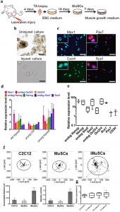
Figure 1
IMuSCs display stemness and exhibit improved migration ability.
srep17355-f1
(a) Schematic of iMuSCs isolation method from injured murine TA muscles. (b) Bright field images of uninjured and injured cultures. 3 days after the cell isolation no cells appeared in the control uninjured cultures, but iMuSCs were present in the injured cultures. 7 days after cell isolation, the proliferation of iMuSCs was apparent. Scale bar = 10 μm. (c) Msx1 (green), Pax7 (red), Cxcr4 (green), and Sca1 (red) expression of iMuSCs. Nuclei were stained with DAPI (blue). Scale bar = 100 μm. (d) qPCR analysis of whole biopsied TA muscles, and (e), fresh isolated iMuSCs. (f) Single cell migration pathways of iMuSCs, and the control C2C12 and MuSCs. The migration paths of 20 individual cells from different experimental groups captured in a time-lapse motility assay. Data was pooled from 3 independent experiments. Graphs show the calculated accumulated distance and velocity of the cells. Data are represented as the mean ±SEM of 60 individual cells from 3 biological replicates. **P < 0.01. (g) qPCR analysis of β-Catenin, E-Cadherin, M-Cadherin, N-Cadherin expression of iMuSCs. Data are represented as the mean ±SEM of 5 biological replicates.
図1
iMuSCsは幹細胞性を示し、優れた移動能力を発揮する。
(a)傷害マウスTA筋からのiMuSCs単離方法のシェーマ。(b)非傷害マウス由来細胞培養と傷害マウス由来細胞培養。細胞単離後3日目、非障害マウス由来細胞培養からは何も得られなかったが、傷害マウス由来細胞の培養からiMuSCsが出現した。細胞単離後7日目、iMuSCsの増殖が鮮明に認められた。Scale bar = 10 μm。(c) iMuSCsにおけるMsx1(緑)、Pax7(赤)、Cxcr4(緑)、Sca1(赤)の発現。核は DAPI(青)で染色した。Scale bar = 100 μm。(d)TA筋全生検のqPCR解析、及び(e)新鮮単離iMuSCsのqPCR解析、(f)iMuSCsの単細胞遊走路、及びコントロールC2C12、MuSCsの単細胞遊走路。低速度撮影運動実験において捉えられた異なる実験グループ由来の20個の細胞の移動道程。3回の独立した実験からデータを収集した。グラフは、細胞の算出後の移動距離と速度を示す。データは、3回反復実験から得られた異なる60個の細胞の平均±SEMで表わした。**P < 0.01。(g)iMuSCsにおけるβカテニン、E-カヘドリン、M-カヘドリン、N-カヘドリン発現のqPCR解析。データは、5回反復実験の平均±SEMで表わした。
カテゴリー: 最新医学知識 | iMuSCs, #10(1) はコメントを受け付けていません

iMuSCs, #9(3)

Methods, part3
Karyotype analysis
Karyotypes were determined using conventional G-Banding analysis at the Texas Children’s Hospital Cytogenetic Core Lab, Houston, TX, USA.
Population doubling assay and histochemical staining for alkaline phosphatase
To estimate the growth of cultures, a standard protocol was followed for population doubling assays. Briefly, 5000 iMuSCs were plated on collagen type IV-coated 6-well plates in growth medium for four days, and every day the well content was collected and counted. The approximate population doubling time (PDT) was determined as: , where, t = time period (24 h), N0 = initial cell number, Nt = number of cells at the time period.
Histological staining for alkaline phosphatase activity was carried out using a commercially available kit (Sigma-Aldrich, USA) following the manufacturer’s instructions.
Quantitative real-time PCR
Total RNA was isolated using the RNeasy Plus Mini Kit (Quiagen, USA), and cDNA was synthesized from 1 μg of RNA via a High Capacity RNA-to-cDNA Kit (Life Technologies, USA) following the manufacturer’s instructions. Gene expression was analyzed by quantitative real-time PCR (qPCR) using MyiQ real time PCR System (Bio-Rad, USA). The applied primers (Supplementary Table 3) were designed by Oligo software (Oligo Perfect Designer, Invitrogen, USA). Reactions were measured in duplicates using custom 2× Syber Green Master Mix based on the hot-start Jumpstart Taq DNA Polymerase enzyme (Sigma, USA). The amplification was done for 40 cycles (95 °C 20 sec, 60 °C 20 sec, 72 °C 40 sec). To verify the PCR products, melting curves and negative controls were carried out in each reaction. Relative quantification of mRNA was determined by the ΔΔCt method (2−ΔΔCt formula)19 by using the expression profile of the corresponding control samples as reference.
Data processing and statistical analysis
Prism 6.0 (GraphPad Software, USA) was used for data plotting, non-linear regression and statistical analysis. Data are given as mean ± S.E.M. Comparisons between two groups were performed by using Student’s t-test assuming two-tailed distribution, and unequal variances. For multiple comparisons, ANOVA or Kruskal-Wallis test was applied. Statistical significance was considered at p < 0.05.
 実験方法(3)
 核型分析
核型は、テキサス子ども病院細胞遺伝学コア研究所(ヒューストン、TX、USA)の標準的Gバンディング解析を使って決定した。
 細胞数倍加アッセイとアルカリフォスファターゼの組織化学的染色法
培養細胞の成長度合いを検討するために、標準的プロトコールにより、細胞数倍加アッセイを行なった。簡単に、5000個のiMuSCsを、4型コラーゲンコーティング6穴プレートに撒き、成長培養液を用い4日間培養し、細胞を毎日回収して細胞数を算出した。倍加時間の算出法は次の通り:Nt/N0=2(t/PDT)、t=時間間隔(24時間)、NO=その時間間隔における細胞数。アルカリフォスファターゼの組織染色は売られているキット (Sigma-Aldrich, USA)を使用して、添付文書に従い行なった。
 量的リアルタイムPCR
全RNAはRNeasy Plus Mini Kit (Quiagen, USA)より分離、またcDNAはHigh Capacity RNA-to-cDNA Kit (Life Technologies, USA)を用い、添付文書に従って、1μgのRNAから合成した。遺伝子発現は、MyiQ real time PCR System (Bio-Rad, USA)を使い、量的リアルタイムPCRによって分析した。使用したプライマー(補足テーブル3)は、Oligo software社 (Oligo Perfect Designer, Invitrogen, USA)により作成して頂いた。反応は、hot-start Jumpstart Taq DNAポリメラーゼ(Sigma, USA)に基づいて、custom 2× Syber Green Master Mixを使って測定した。増幅は40サイクル(95℃で20秒、72℃で40秒)行なった。PCR産物量を確認するため、融解曲線とコントロールをそれぞれの反応において行なった。mRNAの相対量は、参考文献にあるように対応対照サンプルの発現状況を使って、ΔΔCt法(2−ΔΔCt formula)19によって判定した。
データ処理と統計学的解析
データの描出、非直線回帰、統計学的解析は、Prism 6.0 (GraphPad Software, USA)解析ソフトを用いて行なった。データは平均±S.E.M.にて算出した。2グループ間の比較は、両側分布を仮定しStudentのt-検定及び不等分散で行なった。多重比較に関しては、ANOVAまたはKruskal-Wallis検定を適応し施行した。統計学的有意差は、p < 0.05において有意とした。


カテゴリー: 最新医学知識 | iMuSCs, #9(3) はコメントを受け付けていません

iMuSCs, #9(2)

Methods, part2
In vitro differentiation
In vitro embryoid body (EB) formation of iMuSCs was induced by applying the routinely used ‘hanging-drop’ technique6,16. Single cell suspensions of iMuSCs (3.75 × 104 cells/ml) were placed as 20 μl micro-drops of ESGRO Complete PLUS Clonal Grade Medium (Millipore, USA) on the lids of 100 mm dishes. Bottom plates of the dishes were filled with sterile PBS to avoid desiccation of samples. Lids were then placed on the bottom plates for 4 days to achieve hanging drop cultures. During this time, single iMuSCs formed EBs, which were harvested and further cultured in suspension for 3 additional days. For spontaneous in vitro differentiation, EBs were cultured on collagen type IV-coated 24-well plates in EB Medium [DMEM supplemented with 20% FBS, 2 mM Glutamax, 1% non-essential amino acids, and 1% antibiotics; all from Gibco (USA)] for 2 weeks with medium changed every other day. Additionally, multiple differentiations of iMuSCs were assessed by myogenic and osteogenic differentiation assays following standard protocols.
Singe cell migration
Target cell populations and control cells (MuSCs and C2C12 cells) were plated separately on collagen type IV-coated 6-well plates, 10,000 cells/well, in growth medium, 48 hours prior to the time-lapse experiment. Time lapse images were acquired with an Andor IXon3 885 EMCCD camera (Andor, USA) on an Olympus IX-81 (Olympus, USA) microscope fitted with a microscope enclosure (Precision Plastics, USA), and images of single cell migration were taken for six hours at three minute intervals; three different fields/well were chosen for the recording. Proper environmental conditions were maintained in a micro incubator at 37 °C with 5% CO2. A series of images were analyzed using NIH ImageJ analysis software to track the centroid positions (x, y) of cell nuclei (which were assumed to be the representations of cell-bodies). Migration paths were plotted and analyzed by the Chemotaxis and Migration Tool v2.0 from Ibidi. Net translocation distance was measured as the distance between the starting point and the end point of cells after six hours. Migration speed was calculated as total length of the migration path during the six-hour period.
Immunohistochemistry
Samples were fixed with 4% paraformaldehyde (Sigma, USA) for 20 minutes at room temperature. After permeabilization with 0.2% Triton X-100 (Sigma, USA) for 30 minutes, nonspecific binding of antibodies was blocked for one hour with 10% BSA and 5% HS (Sigma, USA) in PBS, at room temperature. The primary antibodies (Supplementary Table 2) were applied overnight at 4 °C. After the overnight incubation, the cells were incubated for one hour at room temperature with the appropriate fluorescence-conjugated secondary antibodies (Extended Data Table 2). The nuclei were revealed using 4′, 6′-diamidino-2-phenylindole (DAPI) staining (Sigma, USA), and fluorescent microscopy (Nikon) was used to visualize the results. Quantitative image analysis was performed using the NIH ImageJ Software.
実験方法(2)
In vitroでの分化誘導
In vitroでのiMuSCs胚様体(EB)形成は、頻用されている「懸滴培養」技術を応用することにより誘導した。iMuSCs (3.75 × 104 cells/ml)単細胞浮遊液を、20μlずつ微小滴下し、ESGRO Complete PLUS Clonal Grade培養液(Millipore, USA) を満たしたmicro-drops100 mm培養皿の蓋に撒いた。サンプルの乾燥を避けるために培養皿の底部を滅菌PBSで満たした。懸滴培養を実施するため、培養皿の蓋部を4日間培養皿の底部の上に置いた。培養4日間で、単細胞iMuSCsは胚様体を形成した。それを回収し、さらに3日間、浮遊状態で培養を続けた。自発的in vitro分化誘導のため、胚様体は、EB用培養液[DMEMに添加20% FBS、2mMグルタミン酸、1%非必須アミノ酸及び1% 抗体;すべてGibco (USA)より購入]を用いて4型コラーゲンコーティング24穴培養プレートで2週間培養した。培養液は2日ごとに交換した。加えて、iMuSCsの多能性を検討するため、標準的なプロトコールに従って筋源性及び骨源性分化実験を行なった。
単細胞遊走能
標的細胞集団とコントロール細胞(MuSCs及びC2C12細胞)をそれぞれ10,000細胞数/ 穴の濃度で、4型コラーゲンコーティング6穴培養プレートに撒いた。低速度撮影に先立ち、成長培養液を用い48時間培養した。低速度撮影画像は、顕微鏡格納装置(Precision Plastics, USA)付きオリンパスIX-81(Olympus, USA)顕微鏡を装着したAndor IXon3 885 EMCCDカメラ(Andor, USA)から得た。単細胞遊走の画像は、3分間隔で6時間撮影した。1つのプレートのウエル(穴)当たり3つの異なるフィールドを記録用として選択した。適切な培養環境を維持するために、マイクロインキュベーター(培養器)は、37°C、5% CO2下で管理した。一連の画像は、NIH ImageJ解析ソフトウェアで分析し、細胞核の重心(細胞体の代表と仮定した)の位置 (x, y) を追跡した。遊走経路は、Ibidiから提供されたChemotaxis and Migration Tool v2.0にょって分析し、プロットした。正味の移動距離は、開始点から6時間後の終了点の距離として算出した。遊走スピードは6時間の全遊走距離として計算した。
免疫化学的検討
サンプルは4%ホルムアルデヒド (Sigma, USA)を用いて、20分間、室温で固定した。0.2%トリトンX-100 (Sigma, USA)で30分間透過処理後、10% BSAと5% HS (Sigma, USA) 添加PBSを用い室温で1時間、非特異的抗体を結合させた。一次抗体(補足テーブル2)は、4°Cで一晩反応させた。一晩放置した後、細胞は、蛍光抱合2次抗体を用いて室温で1時間反応させた。核は、4′, 6′-ジアミジノ-2-フェニールインドール(DAPI)(Sigma, USA) 染色法で染色し、蛍光顕微鏡((Nikon)のより画像を描出した。量的イメージ解析にはNIH ImageJ Softwareを使用した。


カテゴリー: 最新医学知識 | iMuSCs, #9(2) はコメントを受け付けていません

iMuSCs, #9(1)

Methods, part
Animal studies
The animal experiments and all experimental protocols were approved by the Center for Laboratory Animal Medicine and Care at The University of Texas Health Science Center at Houston (protocol No: AWC-13-134). All methods were carried out in accordance with the approved relevant guidelines and regulations. Mouse strains utilized in this study: C57BL/6J, BALB/c, SCID-beige, and mdx/SCID were purchased from the Jackson Laboratory, USA.

Cell isolation and maintenance
Mouse iMuSCs were isolated from the injured TA muscles of C57BL/6J (3–8-week-old female; Jackson Lab, USA) mice four days after laceration injury, while control MuSCs were isolated from uninjured TA muscles. The iMuSCs were separately cultured in ESGRO Complete PLUS Clonal Grade Medium (Millipore, USA) on 12-well tissue culture plates (Corning, USA) for 3 weeks. The medium was then replaced with normal muscle growth medium [Dulbecco’s Modified Eagle’s Medium (DMEM) supplemented with 20% Fetal Bovine Serum (FBS), 10% Horse Serum (HS), 1% chicken embryo extract (CEE; Accurate Chemical Co., UK), and 1% Penicillin-Streptomycin antibiotics; unless otherwise mentioned, all from Gibco, USA] and iMuSCs were further cultured and expanded on collagen type IV-coated flasks at 5% CO2 at 37 °C. C2C12 primary mouse myoblasts (purchased from ATTC, USA) and MuSCs were used as controls and were cultured on collagen type IV-coated flasks in growth medium in 5% CO2 at 37 °C. Characterization of iMuSCs was performed by applying standard in vitro and in vivo assays.
Generation and analysis of chimeric mice
Undifferentiated single cells of β-gal- and GFP-pre-labelled iMuSCs were transferred into blastocysts of BALB/c mice (Jackson Lab, USA) by microinjection following standard procedures8. Pregnant mice were sacrificed and E14 embryos were collected and dehydrated in a serial dilution of 30, 20 and 10% sucrose and then fixed in 4% paraformaldehyde (Sigma, USA) and embedded in paraffin. Sections were stained with anti-GFP antibody, counterstained with eosin and visualized by fluorescent microscopy (Nikon).
Teratoma formation assay
IMuSCs were suspended at 1 − 2 × 106 cells/ml in Phosphate Buffered Saline (PBS). SCID-beige mice (Jackson Lab, USA) were anesthetized with diethyl ether and injected with 500 μl of the cell suspension subcutaneously into the dorsal flank. Seven weeks after the cell injection, tumours were surgically dissected from the mice. Samples were weighed, fixed in 4% formaldehyde, and embedded in paraffin. Sections were stained with hematoxylin and eosin.
 実験方法(1)
動物実験
動物実験とすべての実験手順は、ヒューストン・テキサス大学テキサス健康科学センター内動物医学ケアセンター研究所(the Center for Laboratory Animal Medicine and Care at The University of Texas Health Science Center at Houston)のプロトコール第AWC-13-134に従って了承された。すべての方法は、承認済ガイドラインと規則に準じて施行された。当該研究において使用されたマウスは次の通り:C57BL/6J、BALB/c、SCID-beige、およびmdx/SCIDの4種で、アメリカ、ジャクソン研究所(Jackson Laboratory, USA)より購入した。
細胞の単離と培養
iMuSCsは、C57BL/6Jマウス(3~8週齢の雌をJackson Lab, USAより購入)を用いて、傷害後4日目のTA筋から単離した。一方、対照MuSCsは、非傷害TA筋から単離した。iMuSCsは、培養液ESGRO Complete PLUS Clonal Grade Medium (Millipore, USA)、及び12穴培養プレート(Corning, USA)を用いて、3週間培養を継続した。その後培養液を正常筋成長培地[10%馬血清(HS)1%鶏胎抽出液(CEE、Accurate Chemical Co., UK)1%ペニシリン-ストレプトマイシンをそれぞれ添加したDulbecco’s Modified Eagle’s Medium (DMEM)、特に言及がなければ、すべてGibco, USAより購入]で置換した。そして、iMuSCsはさらに培養を続け、5% CO2、 37 °Cの下で、4型コラーゲンをコーティングした培養フラスコで継代した。C2C12初代マウス筋芽細胞(ATTC, USAより購入)とMuSCsはコントロールとして使用し、コラーゲン4型をコーティングフラスコを用い、5% CO2、37 °Cの条件で培養した。iMuSCsの性質は、標準的in vitro及びin vivo実験を行ない検討した。
キメラマウスの発生と分析
β-galとGFPで予めラベルした未分化単細胞iMuSCsを、標準的なマイクロインジェクション法により、BALB/cマウス(Jackson Lab, USA)由来胚盤胞へと移入した。妊娠マウスを犠牲にし、E14胎児を取り出した。得られた胎児は、30、20、10%ショ糖希釈液にて脱水し、4%パラホルムアルデヒド(Sigma, USA)にて固定後パラフィン包埋した。切片は抗GFP抗体で染色、エオジンを用いて対比染色し、蛍光顕微鏡(Nikon)で可視化した。
奇形腫作成実験
リン酸バッファー生食(PBS)を用い、1-2×10の6乗個/mlのiMuSCs細胞の浮遊液を作成した。ジエチルエーテルを使って、SCID-beigeマウス(Jackson Lab, USA)を麻酔し、500 μlの細胞浮遊液を背側面の皮下に注射した。細胞浮遊液を注射してから7週間後、マウスから腫瘍を外科的に摘出した。得られたサンプルの重量を測定、4%ホルムアルデヒドで固定し、パラフィン包埋した。切片はへマトキシリン・エオジン染色した。


カテゴリー: 最新医学知識 | iMuSCs, #9(1) はコメントを受け付けていません